Novel Method for Counting Nanoparticles in Biological Tissues
Technology description
| The name of the technology: | Novel Method for Counting Nanoparticles in Biological Tissues |
|---|---|
| Challenge: | Numerous techniques for sensitive biomarker imaging in biological tissues employ chemical or nanoparticle tags. Electron microscopy can detect a single nanoparticle and offers high spatial resolution, but it is rather time-consuming and does not allow multiplexing, i.e., simultaneous detection of multiple biomarker types. Fluorescence microscopy suffers from autofluorescence, and its multiplexing capability is limited due to the broad absorption and emission spectra of tags. Imaging mass cytometry uses a set of nanoparticle tags that enormously improve sensitivity and multiplexing compared to fluorescent techniques, yet it does not offer detection at the single-molecule level. |
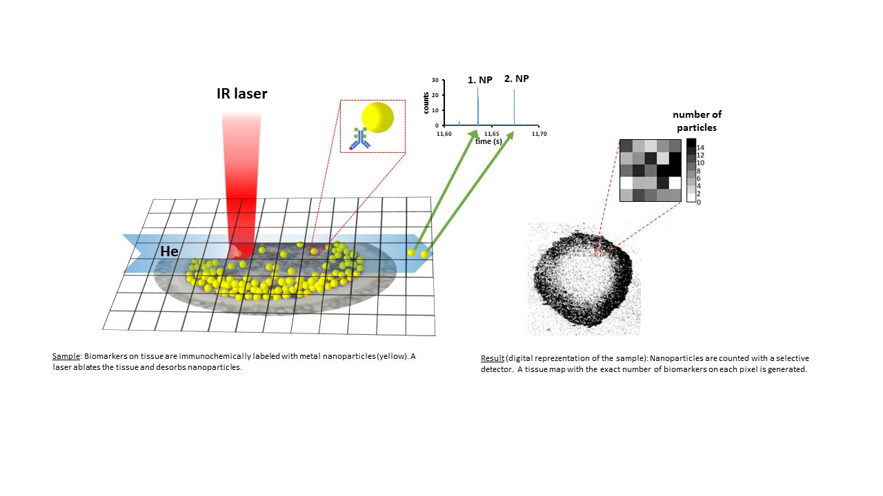
| Description: |
Our laser ablation technique employs an infrared laser and a simple ablation cell to count metal nanoparticle tags on biological tissues or other organic substrates. Unlike the conventional laser ablation systems (imaging mass cytometry), where nanoparticles disintegrate during the ablation process, we can desorb intact nanoparticles and count them. The technique is demonstrated in monitoring proliferating cells in 3D aggregates of human colorectal carcinoma cells. Precise counting of the tags on each pixel generates sharp distribution maps of a proliferation biomarker in the tissue. Advantageously, signals from regions outside the tissue are strongly suppressed. The technique is not limited to biological tissues and can enumerate nanoparticle tags selectively bound to immunosorbents. It can be used for quantitative analyses in an analogy to enzyme assays, such as ELISA, etc. Benefits: - nanoparticle detection and counting with ultimate, single nanoparticle sensitivity - an analogy to single photon (digital) counting vs. proportional (analogous) light measurement - virtually unlimited multiplexing - more sensitive alternative to confocal microscopy and imaging mass cytometry Applications: - tissue imaging - immunoassays |
| Commercial opportunity: | The technology has the potential to enhance the imaging scope of mass spectrometers. |
| IP protection status: | Patent pending - PCT/CZ2023/050031 |
| Development status: |
Phase 2Corresponds with TRL 3 and TRL 4 Feasibility study. There is a realistic design of the technology and the initial tests in the laboratory are leading to the specification of the technology requirements and its capabilities.
|
| Partnering strategy: | Collaboration licensing |
| More information: | We are searching for potential partners in development (financial support of further development), commercialization (licensing), and exploration of further application potential. |
| Images: |
|
| Categories: | Chemistry Chemical and bioengineering |
| Owner of a technology: | Masarykova univerzita |